Orbital Fracture
The human eye rests securely within a bony cavity known as the orbit, which provides protection and structural support. However, trauma or forceful impact to the face can cause the thin bones surrounding the eye to crack or break—a condition known as an orbital fracture. This injury not only affects the bones but can also damage the muscles, nerves, and tissues around the eye, leading to pain, swelling, and vision problems if left untreated.
The orbit is a complex structure composed of seven bones that cradle and protect the eyeball. It also houses muscles, nerves, and fat that allow smooth eye movement and cushioning. When a strong blow—such as from a fall, car accident, sports injury, or assault—hits the eye or surrounding area, the thin bones of the orbital floor or wall can fracture. Depending on the impact, the break can range from a simple crack to a displaced bone fragment.
Types of Orbital Fractures
Orbital fractures are classified based on their location and the extent of damage:
- Orbital Rim Fracture: This involves a break in the outer edge of the eye socket, often caused by severe trauma such as motor vehicle accidents. Since the orbital rim is thick and strong, such fractures often occur along with other facial bone injuries.
- Blowout Fracture: A blowout fracture occurs when a sudden impact increases pressure within the orbit, causing the thin floor or inner wall to break while the outer rim remains intact. Soft tissues like muscles and fat can become trapped in the fracture, leading to restricted eye movement and double vision.
- Orbital Roof Fracture: More common in children, this type affects the upper portion of the orbit. It may be associated with head injuries and requires careful neurological assessment.
- Medial Wall Fracture: The fragile inner wall of the orbit can break, often causing air from the sinuses to enter the surrounding tissues—a condition known as subcutaneous emphysema.
Common Causes of Orbital Fractures
- Blunt trauma from sports injuries (like baseball, boxing, or cricket)
- Motor vehicle accidents involving facial impact
- Falls, especially in the elderly
- Physical assault or violence
- Industrial or workplace injuries involving heavy objects
The severity of the fracture depends on the force and direction of the impact.
Symptoms of an Orbital Fracture
The symptoms can vary depending on which bones and tissues are affected, but the most common signs include:
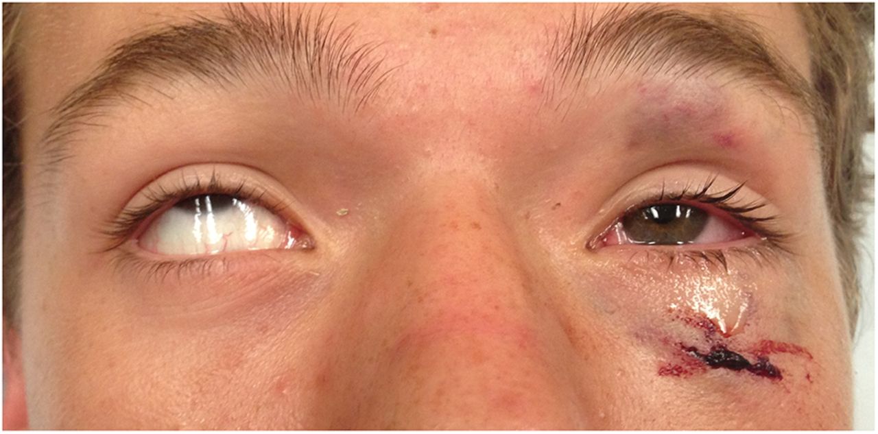
- Swelling and bruising around the eye
- Pain and tenderness when moving the eye
- Double vision (diplopia)
- Decreased eye movement or a feeling of the eye being “stuck”
- Sunken or protruding eyeball (enophthalmos or proptosis)
- Numbness around the cheek, nose, or upper lip (due to nerve injury)
- Blood in the white of the eye (subconjunctival hemorrhage)
- Blurred or decreased vision
- Crepitus (a crackling sound under the skin caused by trapped air)
In severe cases, there may also be eye misalignment or vision-threatening complications such as optic nerve damage.
Diagnosis
A thorough eye and facial examination is crucial for accurate diagnosis. The ophthalmologist or maxillofacial surgeon will perform several assessments, including:
- Visual Acuity Test: To evaluate any loss of vision.
- Eye Movement Examination: To detect restriction or muscle entrapment.
- Palpation: To feel for deformities, tenderness, or air pockets under the skin.
- CT Scan (Computed Tomography): The most reliable imaging test to determine the fracture’s location, size, and involvement of surrounding tissues.
- X-rays: Sometimes used as an initial assessment, though less detailed than CT scans.
Treatment Options for Orbital Fractures
Treatment depends on the severity of the fracture, symptoms, and whether there’s damage to the eye or surrounding tissues.
1. Conservative (Non-Surgical) Treatment
Minor fractures that don’t affect vision, eye position, or muscle movement can often be managed without surgery. Treatment includes:
- Cold Compresses: To reduce swelling.
- Pain Relievers: To control discomfort.
- Antibiotics: To prevent sinus or orbital infections.
- Avoiding Nose Blowing: Prevents air from entering the orbit through damaged sinus walls.
- Observation and Follow-up: Regular check-ups to monitor healing and ensure no delayed complications.
Most small fractures heal on their own within a few weeks with careful management.
2. Surgical Treatment
Surgery is recommended in the following situations:
- Double vision that doesn’t improve with time
- Significant displacement of bones or trapped muscles
- Sunken or misaligned eyeball (cosmetic or functional concern)
- Large fractures involving more than half of the orbital floor
- Evidence of optic nerve compression or vision loss
Surgical Procedure: The goal of surgery is to restore the normal structure of the orbit, release trapped tissues, and rebuild the bone if needed. The procedure typically involves:
- Accessing the fracture through a small incision inside the eyelid or under the lower lash line.
- Gently repositioning displaced tissues and muscles.
- Placing a thin implant or mesh plate to reconstruct the orbital floor or wall.
- Closing the incision carefully to minimize scarring.
Modern surgical techniques, including endoscopic and minimally invasive methods, allow for precise repair and faster recovery.
Recovery and Postoperative Care
Recovery time varies depending on the severity of the fracture and whether surgery was performed. General care recommendations include:
- Rest and avoid strenuous activities for several weeks.
- Use prescribed eye drops or ointments to prevent infection and dryness.
- Apply cold compresses during the first few days to minimize swelling.
- Elevate the head while sleeping to reduce fluid buildup.
- Avoid nose blowing for at least two weeks after surgery.
- Attend all follow-up appointments to monitor healing and eye alignment.
Most patients experience significant improvement in appearance, eye function, and comfort within weeks, though complete recovery can take several months.
Possible Complications
If not treated properly, orbital fractures can lead to long-term complications, including:
- Persistent double vision
- Sunken eye appearance (enophthalmos)
- Chronic pain or numbness around the cheek and eyelid
- Vision loss due to optic nerve damage
- Scarring or deformity of the eye socket
Prompt diagnosis and treatment greatly reduce the risk of these outcomes.
Prevention
While not all accidents can be prevented, certain precautions can minimize the risk of orbital fractures:
- Always wear protective eyewear during contact sports or high-risk activities.
- Use seatbelts and airbags while driving.
- Take care on wet or uneven surfaces to prevent falls.
- Use helmets and safety gear in hazardous work environments.